Why Accurate Kidney Biomarkers Matter
Chronic kidney disease (CKD) now affects roughly 9 % of the world’s population—about 700 million people—and one‑third of these cases reside in China and India. In India, rising diabetes and hypertension have driven CKD prevalence to 8‑10 % of adults, making early detection a public‑health priority. Traditional renal markers—serum creatinine, blood urea nitrogen (BUN) and the derived eGFR—are limited. Creatinine levels are confounded by muscle mass, diet, age, sex and tubular secretion, so they may stay normal until 50 % of filtration capacity is lost. BUN is further influenced by protein intake, liver function and catabolism, reducing its specificity for renal injury. Clinical biochemistry laboratories, such as Agam Diagnostics in Madurai, address these gaps by offering standardized, automated assays for both conventional and emerging biomarkers. Combining serum creatinine with cystatin C improves eGFR precision, while urinary markers (e.g., albumin‑to‑creatinine ratio, KIM‑1, NGAL) detect glomerular or tubular damage weeks before creatinine rises. Rapid, high‑throughput testing and free home collection enable timely risk stratification, allowing clinicians to intervene early and slow CKD progression.
Traditional Kidney Function Tests: Creatinine, BUN, and eGFR
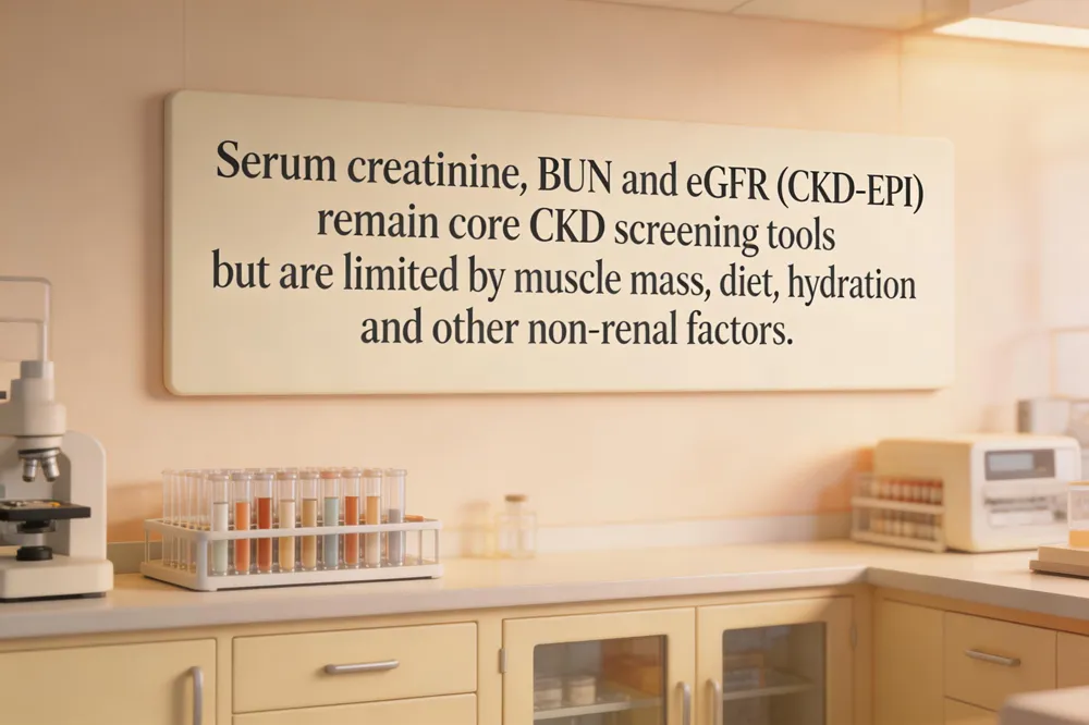
Serum creatinine remains the most widely used biomarker for estimating kidney filtration, but its concentration is heavily influenced by non‑renal factors such as muscle mass, diet, age, and sex. Because creatinine is produced from muscle creatine metabolism, individuals with low muscle mass (elderly, malnourished, or chronic illness) may have deceptively low levels, while high‑protein diets or certain medications can raise it independently of glomerular function.
Blood urea nitrogen (BUN) reflects the amount of urea in the bloodstream, a waste product of protein catabolism. Although BUN rises when renal clearance falls, it lacks specificity: dehydration, high protein intake, liver disease, and gastrointestinal bleeding can also elevate BUN, limiting its reliability as a stand‑alone indicator of kidney injury.
Estimated glomerular filtration rate (eGFR) translates serum creatinine (or cystatin C) into a functional measure of filtration using demographic variables. The CKD‑EPI equation is currently preferred over the older MDRD formula because it provides more accurate estimates across a broader range of GFR values, especially >60 mL/min/1.73 m². Some laboratories also report eGFR derived from combined creatinine‑cystatin C equations, which improve precision in diverse populations.
KDIGO staging categorises chronic kidney disease based on eGFR: Stage 1 ≥90, Stage 2 60‑89, Stage 3 44‑59, Stage 4 15‑29, and Stage 5 <15 mL/min/1.73 m². These stages guide clinical decision‑making, referral thresholds, and monitoring frequency, emphasizing the need for accurate e[FR calculation and awareness of the limitations of creatinine and BUN alone.
Cystatin C – The Muscle‑Mass‑Independent Filtration Marker
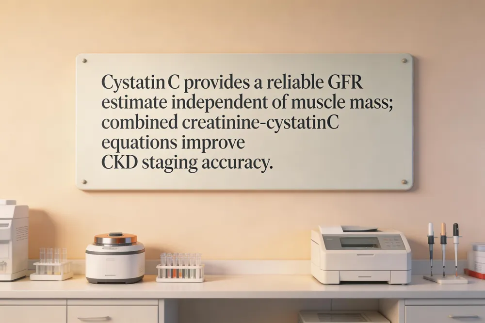
Cystatin C (CysC) is a 13‑kDa cysteine‑protease inhibitor produced at a constant rate by all nucleated cells. Because it is freely filtered at the glomerulus and neither re‑absorbed nor secreted by the renal tubules, its serum concentration reflects the true glomerular filtration rate (GFR) with minimal physiologic interference. Numerous studies have shown that serum CysC correlates tightly with measured GFR (mGFR) and often outperforms serum creatinine, especially when muscle mass, diet, or age distort creatinine‑based estimates. In elderly patients, individuals with malnutrition, and Asian populations where lower average muscle mass can lead to under‑estimation of kidney function, CysC provides a more reliable assessment of filtration capacity. Modern clinical practice therefore increasingly incorporates CysC into eGFR equations. The CKD‑EPI Cys‑Cr (creatinine‑cystatin C) combined formula , for example, integrates both markers and consistently yields higher precision than either marker alone, reducing bias and improving classification of CKD stages. This dual‑marker approach is now recommended by KDIGO and is widely adopted in automated renal panels, such as those offered by Agam Diagnostics, to support early detection and accurate monitoring of kidney disease.
Urinary Albumin‑to‑Creatinine Ratio (UACR) – Early Glomerular Damage Detector
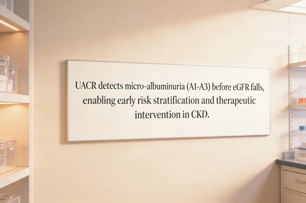
Urinary microalbumin (mALB) is a sensitive early marker of glomerular damage, especially in diabetic and hypertensive nephropathy, and is used in the albumin‑to‑creatinine ratio (UACR) for CKD staging. When the glomerular filtration barrier becomes permeable, low‑level albumin leaks into urine, which can be quantified as the urine albumin‑to‑creatinine ratio (UACR). The KDIGO 2024 guidelines define three albuminuria categories: A1 (UACR < 30 mg/g) considered normal, A2 (30‑300 mg/g) indicating moderate albuminuria, and A3 (> 300 mg/g) reflecting severe albuminuria. These thresholds are independent of eGFR and provide prognostic information beyond filtration rate alone. For risk stratification, KDIGO recommends combining UACR with eGFR: patients with eGFR ≥ 60 mL/min/1.73 m² but A2 or A3 albuminuria are re‑classified to higher CK‑ risk stages because albuminuria predicts faster eGFR decline, cardiovascular events, and mortality. Conversely, a normal UACR can offset a mildly reduced eGFR (60‑89 mL/min/1.73 m²) in staging. Routine measurement of UACR, together with serum creatinine‑ or cystatin‑C‑derived eGFR, therefore enables early detection of glomerular damage, guides therapeutic intensification, and improves long‑term outcomes for patients at risk of CKD.
Emerging Tubular Injury Biomarkers for Acute Kidney Injury (AKI)
![NGAL, KIM‑1, L‑FABP and [TIMP‑2]·[IGFBP‑7] rise within hours of tubular damage, allowing earlier AKI diagnosis and risk prediction.](https://rank-ai-generated-images.s3-us-east-2.amazonaws.com/30d7f2cf-34cb-4bd9-8194-211654acdffc-banner-c670a6ad-35a2-42f0-a519-2f9ab5e5a8ee.webp)
Acute kidney injury (AKI) often develops before any measurable rise in serum creatinine, making early tubular injury biomarkers essential for timely diagnosis and intervention. Neutrophil gelatinase‑associated lipocalin (NGAL) is a 25‑kDa protein that appears in urine and plasma within 2–6 hours of tubular damage, rising rapidly and correlating with injury severity and outcomes in cardiac surgery, sepsis, and critical‑care settings. Kidney injury molecule‑1 (KIM‑1) is a transmembrane glycoprotein expressed almost exclusively on injured proximal tubular cells; it is undetectable in healthy urine and becomes markedly elevated after ischemic or toxic injury, providing a specific signal of proximal tubular dysfunction. L‑type fatty‑acid‑binding protein (L‑FABP) is released from proximal tubular cells during ischemic stress; its urinary concentration rises before changes in serum creatinine and helps differentiate intrinsic tubular injury from prerenal causes. The combination product tissue inhibitor of metalloproteinisms‑2 (TIMP‑2) and insulin‑like growth factor‑binding protein‑7 (IGFBP‑7) reflects G1‑cell‑cycle arrest in stressed tubular cells, and the product [TIMP‑2]·[IGFBP‑7] (commercially known as NephroCheck) has received FDA approval for predicting moderate‑to‑severe AKI within 12 hours in critically ill patients. Together, these markers enable clinicians to detect AKI earlier, stratify risk, and initiate nephroprotective strategies before irreversible loss of glomerular filtration occurs.
Low‑Molecular‑Weight Proteins as Indicators of Tubular Dysfunction
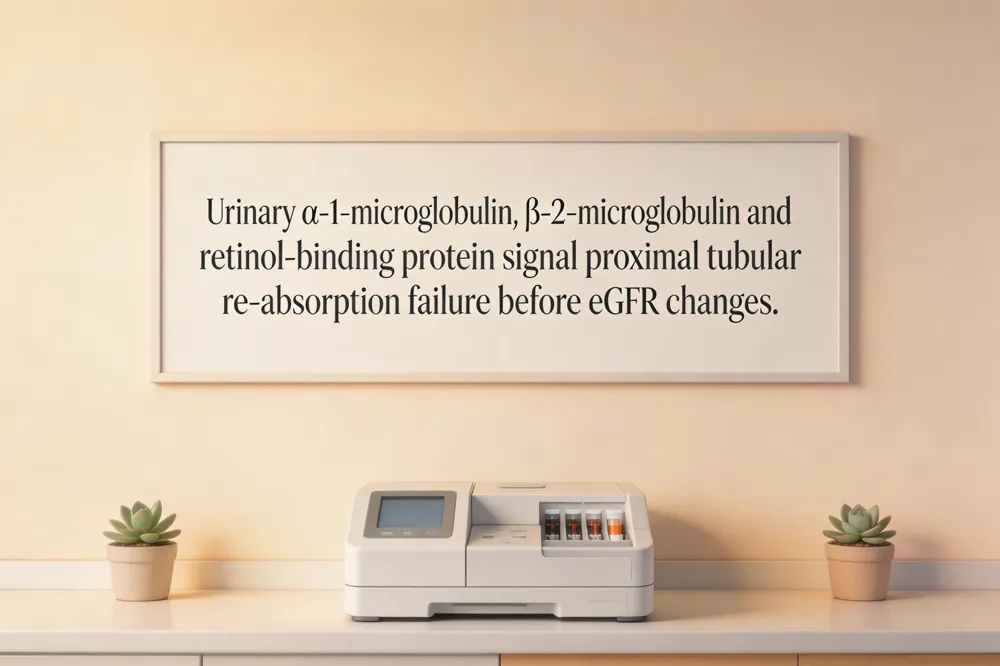
Low‑molecular‑weight proteins (LMWPs) such as α‑1‑microglobulin (A1MG), β‑2‑microglobulin (B2MG) and retinol‑binding protein (RBP) are freely filtered at the glomerulus and normally re‑absorbed by the proximal tubular epithelium. When tubular cells are injured, the re‑absorption capacity diminishes, causing rapid urinary excretion of these proteins—often before overt changes in serum creatinine or eGFR become apparent.
Because their rise reflects proximal tubular damage, LMWPs are valuable for early detection of tubular injury. In diabetic nephropathy, urinary A1MG and RBP increase in the pre‑albuminuric phase, signalling tubular stress that precedes glomerular albumin leakage. Similarly, drug‑induced nephrotoxicity—particularly from aminoglycosides, tenofovir, or contrast agents—can be monitored by serial measurements of B2MG and A1MG, which show elevation within days of exposure and correlate with the severity of tubular impairment.
In clinical practice, incorporating LMWP assays into a renal panel enhances risk stratification for patients with diabetes or those receiving potentially nephrotoxic therapies, allowing earlier therapeutic intervention and potentially slowing progression to chronic kidney disease.
Inflammatory and Fibrotic Biomarkers in Chronic Kidney Disease Progression
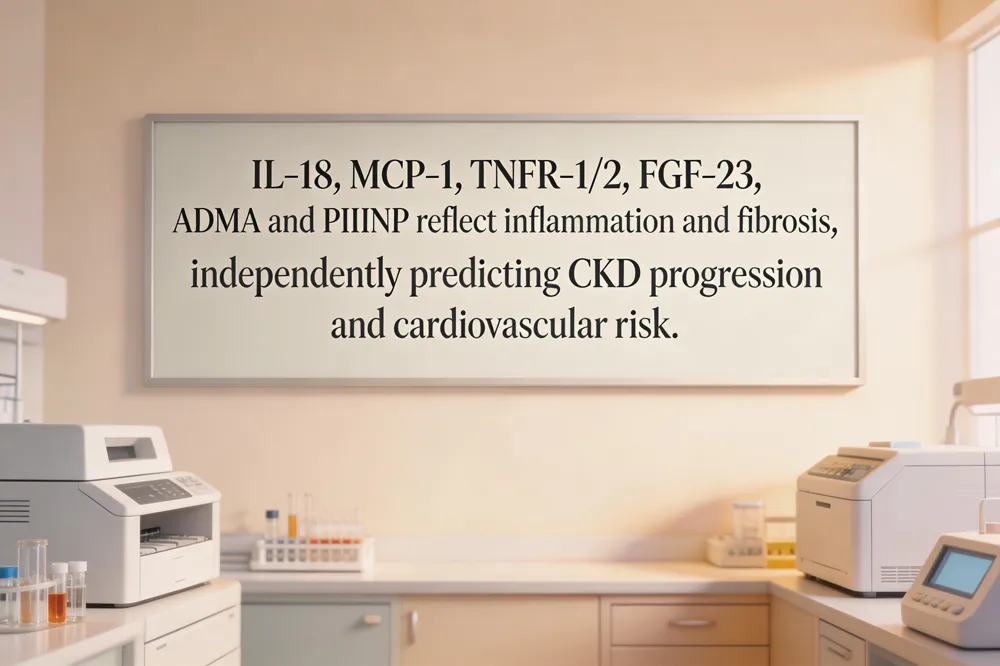
Inflammatory cytokines and fibrotic mediators are increasingly recognised as drivers of CKD progression and cardiovascular complications. Interleukin‑18 (IL‑18) is released from injured tubular cells and rises early in both acute and chronic kidney injury, reflecting innate immune activation and correlating with faster eGFR decline. Monocyte chemoattractant protein‑1 (MCP‑1) is produced by tubular epithelium in response to inflammation; urinary MCP‑1 levels are elevated in diabetic nephropathy and have been linked to accelerated CKD progression. YKL‑40 a chitinase‑like protein involved in tissue remodeling, is also up‑regulated in CKD and predicts both renal outcome and cardiovascular events.
Soluble tumor‑necrosis‑factor receptors 1 and 2 (TNFR‑1, TNFR‑2) circulate as markers of low‑grade inflammation. Large cohort studies have shown that higher plasma TNFR‑1/‑2 concentrations independently predict end‑stage renal disease and cardiovascular mortality, even after adjusting for eGFR and albuminuria.
Fibroblast growth factor‑23 (FGF‑23) rises early in CKD, preceding phosphate and PTH changes, and drives left‑ventricular hypertrophy and vascular calcification. Asymmetric dimethylarginine (ADMA) accumulates as GFR falls, inhibiting nitric‑oxide synthase and contributing to endothelial dysfunction and rapid CKD progression.
Finally, the extracellular matrix remodeling marker procollagen type III N‑terminal propeptide (PIIINP) reflects active fibrosis. Elevated serum PIIINP has been associated with increased interstitial fibrosis, faster eGFR loss, and higher risk of cardiovascular disease, making it a useful surrogate for renal tissue remodeling in CKD patients.
Multiplex Panels and Multi‑omics: The Future of Precision Nephrology
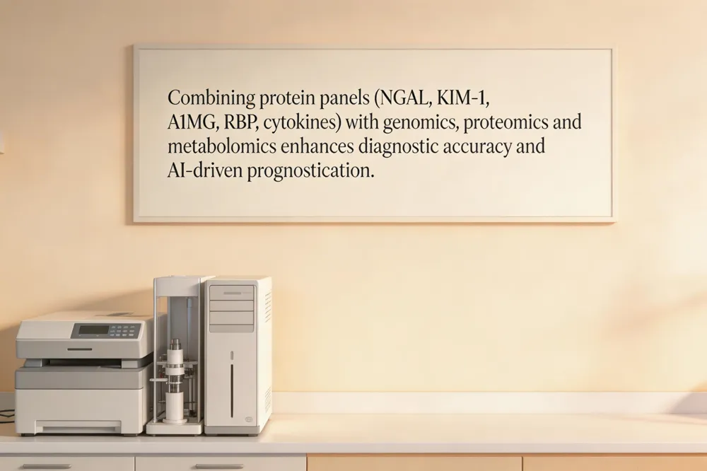
Emerging evidence shows that a single biomarker rarely captures the complex pathophysiology of kidney disease, prompting the development of multiplex panels that combine markers of glomerular filtration, tubular injury, and inflammation. Studies have demonstrated that a panel containing neutrophil gelatinase‑associated lipocalin (NGAL), kidney injury molecule‑1 (KIM‑1), α‑1‑microglobulin (A1MG), retinol‑binding protein (RBP) and inflammatory cytokines such as IL‑18 or MCP‑1 markedly improves diagnostic accuracy for both acute kidney injury (AKI) and chronic kidney disease (CKD) compared with any individual test. The synergistic information from these markers reflects real‑time tubular damage, protein reabsorption failure, and systemic inflammatory response, allowing earlier risk stratification and more precise therapeutic decisions.
Beyond protein panels, integration of genomics, proteomics and metabolomics—collectively termed multi‑omics has accelerated the discovery of novel renal biomarkers. High‑throughput sequencing identifies genetic variants that predispose to CKD, while proteomic profiling uncovers disease‑specific peptide signatures (e.g., CKD273), and metabolomics highlights altered amino‑acid and lipid pathways that precede overt GFR decline. When these data streams are fed into Artificial intelligence and machine learning algorithms, predictive models can synthesize thousands of variables to forecast disease trajectory, dialysis need, or cardiovascular events with unprecedented accuracy.
However, widespread clinical adoption faces hurdles. Standardization of assay methods across platforms remains essential to reduce inter‑laboratory variability, and universally accepted cut‑off values for each biomarker must be established. Regulatory approval pathways for multiplex and multi‑omics tests are still evolving, requiring rigorous validation studies to demonstrate clinical utility and cost‑effectiveness. Overcoming these challenges will be key to translating precision nephrology from research to routine patient care.
Agam Diagnostics: Delivering High‑Quality Kidney Biomarker Testing

Agam Diagnostics in Madurai meets the highest laboratory standards, holding both NABL and ICMR accreditation, which guarantees that all kidney‑function assays—including serum creatinine, cystatin C, neutrophil gelatinase‑associated lipocalin (NGAL), kidney injury molecule‑1 (KIM‑1) and the urine albumin‑to‑creatinine ratio (UACR)—are performed on fully automated analyzers with tight quality‑control procedures. The laboratory’s robotic platforms reduce pre‑analytical variability and deliver reproducible, high‑throughput results. To enhance patient convenience, Agam Diagnostics offers free home‑collection of blood and urine specimens, with most reports generated within six hours of receipt, enabling clinicians to act promptly. All eGFR values are calculated using the CKD‑EPI equations—both creatinine‑based and combined creatinine‑cystatin C formulas—providing standardized, race‑neutral estimates that improve risk stratification for chronic kidney disease. These integrated services ensure accurate, rapid, and reliable kidney‑health assessment for patients across India.
Practical Guidance for Clinicians and Patients
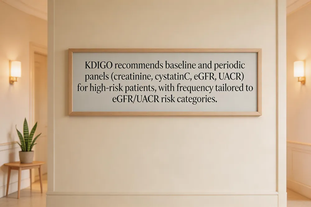
When to order a renal panel – KDIGO recommends routine kidney screening for individuals at high risk of CKD, notably patients with diabetes, hypertension, or those receiving nephrotoxic drugs (e.g., certain antibiotics, NSAIDs, chemotherapy). Early testing should include serum creatinine, cystatin C, estimated GFR (eGFR) using the CKD‑EPI equation, and urine albumin‑to‑creatinine ratio (UACR). In diabetic and hypertensive cohorts, the 2024 KDIGO guideline advises a baseline panel at diagnosis and repeat testing at least annually; more frequent testing (every 3‑6 months) is warranted when eGFR <60 mL/min/1.73 m² or UACR ≥30 mg/g.
Interpretation of eGFR, UACR, and emerging biomarkers – eGFR ≥90 mL/min/1.73 m² is considered normal; 60‑89 mL/min/1.73 m² indicates mild reduction, while <60 signals CKD. UACR <30 mg/g is normal; 30‑300 mg/g reflects moderately increased albuminuria (KDIGO A2) and >300 mg/g signals severely increased albuminuria (A3). Adding cystatin C improves GFR accuracy, especially in low‑muscle‑mass patients. Emerging tubular injury markers such as NGAL, KIM‑1, and L‑FABP can detect early tubular damage and may be used when conventional tests are equivocal or to stratify AKI risk in critically ill patients.
Frequency of monitoring based on KDIGO risk categories – KDIGO risk stratification combines eGFR and UACR. For low‑risk (eGFR ≥60 + UACR <30 mg/g), annual monitoring suffices. Moderate risk (eGFR 45‑59 + UACR 30‑300 mg/g) warrants 6‑month intervals. High risk (eGFR <45 + UACR >300 mg/g) or rapid eGFR decline (>5 mL/min/1.73 m² per year) requires 3‑month follow‑up. Incorporating novel biomarkers (e.g., rising NGAL or KIM‑1) may prompt earlier re‑assessment even when eGFR is stable.
Lifestyle and therapeutic interventions to slow CKD progression – Blood pressure control (<120/80 mm Hg) and optimal glycemic management (HbA1c <7%) remain cornerstone measures. Dietary sodium restriction (<2 g/day), adequate hydration, and avoidance of high‑protein excess reduce renal workload. ACE inhibitors or ARBs are first‑line for proteinuric CKD, decreasing albuminuria and slowing eGFR decline. Smoking cessation, regular aerobic exercise, and weight management further lower cardiovascular and renal risk. Patients should be educated on medication adherence, the importance of regular biomarker testing, and early reporting of symptoms such as edema or changes in urine output.
Economic Impact of Early Biomarker‑Driven Intervention
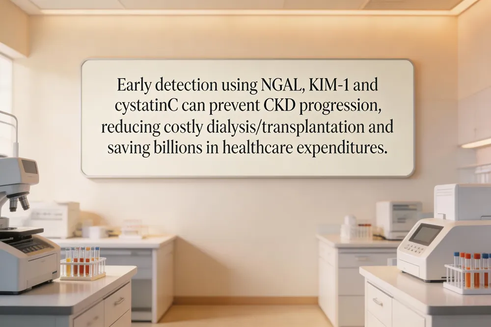
Early detection of kidney injury using emerging biomarkers such as neutrophil gelatinase‑associated lipocalin (NGAL), kidney injury molecule‑1 (KIM‑1) and cystatin C can be highly cost‑effective. [NGAL] and [KIM‑1] rise within hours of tubular damage, allowing clinicians to intervene before serum creatinine and, avoid costly progression to end‑stage kidney disease. [Cystatin C] provides a more accurate estimate of glomerular filtration than creatinine, especially in patients with low muscle mass, reducing unnecessary repeat testing and medication adjustments. In India, where dialysis and transplantation each cost tens of thousands of dollars per year, preventing even a modest proportion of CKD patients from reaching these stages could save billions in health‑care expenditures. National screening programs that incorporate urine albumin‑to‑creatinine ratio (UACR) together with a panel of [NGAL], [KIM‑1] and [cystatin C]—performed in accredited, high‑throughput labs such as Agam Diagnostics—can identify high‑risk individuals early, enable timely lifestyle or therapeutic measures, and lower the downstream burden on dialysis centres and transplant registries. Policymakers should therefore prioritize the inclusion of multi‑biomarker panels in CKD surveillance guidelines, allocate funding for community‑level home collection services, and incentivize laboratories to adopt standardized, automated assays. Such strategies will accelerate early diagnosis, improve patient outcomes, and generate substantial savings for India's public‑health system.
Putting Precision Biomarkers into Everyday Care
Integrating traditional kidney tests—serum creatinine, BUN, eGFR, and urine albumin‑to‑creatinine ratio—with emerging markers such as cystatin C, NGAL, KIM‑1, L‑FABP, and the TIMP‑2 × IGFBP‑7 product creates a multidimensional picture of glomerular filtration, tubular injury, and early inflammation. This synergy improves early detection of acute kidney injury and chronic disease progression far beyond what any single assay can achieve.
Agam Diagnostics in Madurai operationalizes this precision approach by offering fully automated, NABL‑ and ICMR‑accredited panels that combine creatinine‑based eGFR, cystatin C‑derived GFR, and urinary biomarkers (UACR, NGAL, KIM‑1, α‑1‑microglobulin). Free home collection and rapid 6‑hour turn‑around democratize access for patients in remote or underserved regions, ensuring that clinicians receive reliable, standardized results for timely decision‑making.
Looking ahead, AI‑driven dashboards will aggregate longitudinal biomarker data, electronic health records, and imaging to generate personalized risk scores. Expanded panels—including proteomic, metabolomic, and microRNA signatures—will be seamlessly integrated into cloud‑based platforms, enabling continuous, patient‑centric monitoring and proactive therapeutic adjustments before overt renal failure manifests.